- Home
- Departments
Departments Of Intercostal Drainage
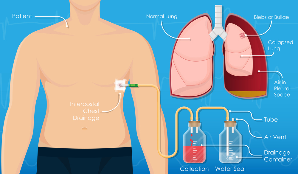
Intercostal-DrainageIntercostal drainage, also known as chest tube insertion or thoracostomy, is a medical procedure used to drain air, fluid, or blood from the pleural space, the thin space between the inner and outer layers of the pleura (the membranes surrounding the lungs). This procedure is commonly performed to treat conditions such as pneumothorax (collapsed lung), pleural effusion (accumulation of fluid), hemothorax (accumulation of blood), or empyema (pus accumulation) in the pleural cavity.
Key Steps of Intercostal Drainage:
Patient Preparation: The patient is positioned comfortably, usually lying on their back or sitting upright. The area where the chest tube will be inserted is cleansed with antiseptic solution, and a sterile drape is placed over the chest to maintain a sterile field.
Anesthesia: Local anesthesia is administered to numb the skin and underlying tissues at the site where the chest tube will be inserted. In some cases, sedation or general anesthesia may be used to help keep the patient comfortable and relaxed during the procedure.
Needle Insertion: A small incision is made in the skin overlying the rib where the chest tube will be inserted. A needle is then inserted through the skin, between the ribs, and into the pleural space under ultrasound or fluoroscopic guidance to minimize the risk of injury to surrounding structures, such as the lung or blood vessels.
Chest Tube Placement: Once the needle is correctly positioned within the pleural space, a chest tube is advanced over the needle and inserted through the incision into the pleural cavity. The chest tube is typically secured to the skin with sutures or adhesive dressings to prevent movement.
Drainage System Connection: The distal end of the chest tube is connected to a drainage system, such as a collection chamber or suction device, which allows air, fluid, or blood to be removed from the pleural space. The drainage system may be set to suction to facilitate drainage, depending on the underlying condition being treated.
Chest Tube Management: The chest tube is monitored regularly to assess the volume and character of drainage and ensure proper function. The tube may be periodically flushed with sterile saline solution to prevent blockage. Chest X-rays may be performed to confirm the placement of the chest tube and monitor the resolution of the underlying condition.
Chest Tube Removal: Once the underlying condition has resolved, the chest tube can be removed. This is usually done by gently pulling the chest tube out through the incision site after ensuring that lung re-expansion and adequate drainage have been achieved. Pressure may be applied to the insertion site to minimize bleeding, and a sterile dressing is applied to cover the wound.
Intercostal drainage is a common and effective procedure for managing various chest conditions requiring drainage of the pleural space. It allows for the removal of air, fluid, or blood, thereby relieving symptoms, promoting lung re-expansion, and preventing complications associated with pleural effusions or pneumothorax. Close monitoring and appropriate management are essential to ensure the safety and success of the procedure.

